Necrotrophic v Biotrophic Pathogenesis
NB: This
link
is to a PDF file which is a good source of further information
· NECROTROPHS…Poorly adapted but
highly virulent pathogens.
1. Bacterial soft-rots. e.g. Erwinia
carotovora.
2. Rapid maceration / killing of host-tissue
· BIOTROPHS…Comparatively less
virulent but more
specialised pathogens.
1. Foliar blights e.g. Pseudomonas
syringae
2. Slower cell-death
3 Very little maceration of the tissue. Subtle interaction
with
host so that nutrients may be extracted over a
long period.
THE NECROTROPHIC PHYTOPATHOGEN:
Erwinia carotovora
Insert Life – cycle
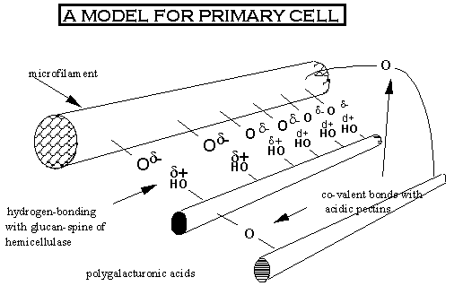
The Barrier :
Plant Cell Wall
1.
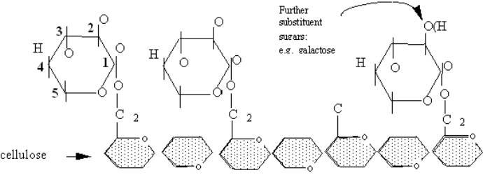
Cellulose:
(the most abundant organic compound in the biosphere...
containing more than 50% organic carbon).
·

unbranched polymer
(1000-1500 Units) of glucose
residues joined at ß-1, 4 linkages.
·
forms
semicrystalline microfibrils: 5-8nm
wide
> 70 cellulose molecules.
·
In
elongating cell are wrapped around the longitudinal axis: 
Cellulose
microfibrils wrapped
Around
the algae,
Chaetomorpha.
2.
 Xyloglucans (1,4)-B-D-glucose polymer, as cellulose
but with xylose at C6.
·
Do
not form microfilaments due to mixed linkages
and side chains.
·
Interaction between xyloglucan polymers are most
likely to be via hydrogen-bonding
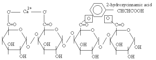
3.
Pectic
polysaccharides
·
Main
polymer : polygalacturonic acid (PGA):
·
a
helical polymer of (1,4)ß-D-galacturonic acid
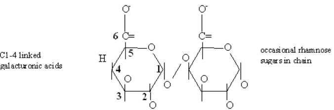
·
Pectic
polymers determine porosity, charge, and pH
and ion balance.
·
PGA is
cross-linked via Ca2+
·
Further
cross-linking occurs via ester linkage with
dihydroxycinnamic acid. :
4. Cell Wall Proteins
·
10%
of cell walls may be composed of proteins
·
Immobilized within the walls via covalent
cross-links
·
Glycine-Rich proteins
(GRPs): glycine
content
may be up to 70% of protein
content of cell wall.
·
Hydroxyproline-rich
glycoprotein (HRGPs)
a.k.a. extensins
·
Not
tightly associated with the cell wall, so
may be considered to be
apoplastic proteins.
·
This
has block of Ser-Pro-Pro-Pro-Pro repeats.
·
Post-translationally
converted to 4-hydroxyprolines
carrying chains (n=1 to 4)
of L-arabinose residues
·
Proposed
to strengthen cell-wall by increasing
X-linking
·
Act
as foci for lignification:
“Virulence”
determinants in Necrotrophic Bacteria
1.
Virulence gene product
· Cellulase
· Xylanases
· Proteases
2.
Pathogenicity gene products
· Pectic lyase and associated enzymes
Pectic lyases- attach
the a-1,-4
glucosidic linkage by
b-elimination produce unsaturated products.
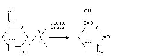
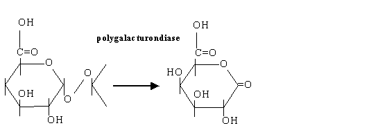
· Polygalacturonidase -cleaves the polymer by hydrolysis
BUT resistance to Erwinia in e.g. potato is associated
with
methylation of the
pectate. An additional enzyme is also
made….
·
Pectic methyltransferases- 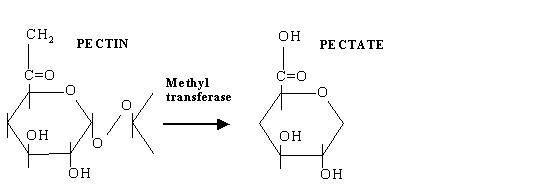
Bad News for the pathogen…
1. The plant has evolved a recognition system to
pectic
fragments.
2. Fragments of between 10-15 residues (degree
of polymerisiation, DP) activate phytoalexins,
PR proteins and lignin biosynthesis.
3. However lower DPs induce further pectinase
activity.
Thus “Battle” over degree of polymerisation.
Quorum Sensing
·
The
mechanism by which bacteria populations sense their size.
·
Vibrio
fischeri,
1.
Symbiont
existing in the light
regions of marine fishes and
squid.
2.
Generates
bioluminescence
through expression of lux
genes
3.
Only active in late exponential
and early stationary phase.
4.
V. fischeri produces an “autoinducer”
Through which-
·
The
bacteria “taste” the medium for the autoinducer to
induce gene expression.
·
Concentration
is proportional to the population size.
5.
N-3-
(oxohexanoyl) homoserine lactone.
A large family of N-acyl homoserine lactones (AHLs) have
now been detected from many species.
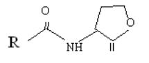
O
6.
10mM
sufficient to activate lux genes
7.
Can
differentiate between free-living (sea) (102 cell/ml)
to light organ
concentrations (1010/11/ml).
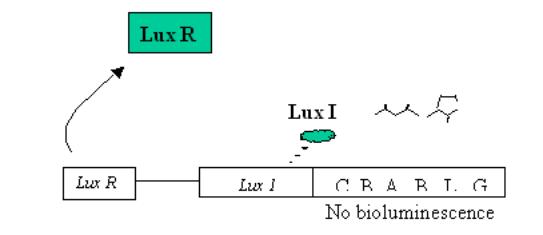
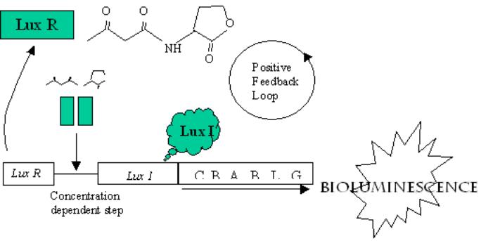
How does quorum
sensing work?
Based on two proteins
(1)
An
AHL synthase (lux I in Vibrio fischeri)
(2)
An
AHL-mediated regulator. (lux R in Vibrio fischeri)
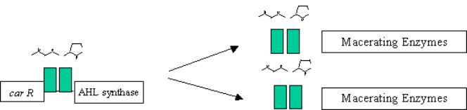
· Vibrio fischeri
At low populations, only a basal amount of AHL is
synthesised
At higher populations, AHL levels
accumulate
·
Erwinia
carotovora