

|
Microbial Growth Website |
|
IBERS: Institute of Biological, Environmental and Rural Sciences |


|
Plate Counting |
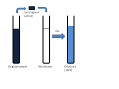
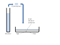
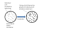
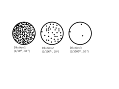
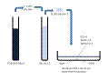
IntroductionOne of the most fundamental microbiological techniques is plate counting which is used to determine the number of viable (i.e. living) cells in a sample. There are several steps to the technique and all must be carried out carefully in order to obtain accurate results. Aseptic technique must be used throughout. Aseptic technique is the term given to a collection of procedures that aim to avoid contamination of the sample. This involves holding tubes at an angle, flaming lids of bottles, using sterile pipettes etc. Plate counting methodStep One: Diluting the sampleDepending on the source of the sample used there might be thousands, millions or even billions of microorganisms per millilitre of sample. This is too many for us to count so we dilute the sample. 1ml of sample is added to 9ml of a suitable diluent (e.g. sterile buffer) (Figure 1a). The sample and diluent are mixed together (Figure 1a). This new sample (Dilution One) has a concentration (number of microorganisms per ml) 1/10th that of the original sample. 1ml of Dilution One is added to another 9ml of diluent to make Dilution Two (Figure 1b). Dilution Two has a concentration 1/10th that of Dilution One and 1/100th that of the original sample. This process is repeated until we have a series of dilutions (Figure 1c). Step Two: Plating the sampleTo find out how many viable cells are in each of our dilutions we need to put some of the sample onto an agar plate. The agar plate is prepared by mixing growth medium with agar and then autoclaving to sterilise. Once the agar has cooled to ~50oC approximately 15ml is poured into a sterile Petri dish and left to set. 0.1ml of sample is pipetted onto the agar surface and spread around using a sterile glass rod. (Figure 2a). We usually put 0.1ml of one of our sample dilutions onto a plate – if we use more than this it can make the plates very wet and if we use less than this it is difficult to spread evenly. This is repeated so that we have 2 or 3 repiclate plates for our original sample and for each of our dilutions (Figure 2b). This is a lot of plates and could get quite expensive and time consuming. We can sometimes use our previous experience to make predictions about the most useful dilutions and make plates just for these. Step 3: Incubating the platesOnce all of the plates have been prepared they are left to dry and then moved to an incubator at a suitable growth temperature for the microorganism being studied. The incubation time depends on the organism and the growth medium but during the incubation, each viable cell that was spread to a discrete position on the agar surface will grow and divide many times to form a visible colony of microorganisms (Figure 3). After the incubation period we can count the colonies to determine how many microorganisms were present in the original sample. Step 4: Counting the coloniesThe plates will have different numbers of colonies depending on the dilution of the sample (Figure 4a). If there are too many colonies it can be impossible or very difficult to count them (Figure 4b). If there is only a small number of colonies it is easy to count them but the results are prone to error (Figure 4c). As a compromise we always aim to count plates with between 30 and 300 colonies (Figure 4d). We record our results noting the dilutions that had between 30 and 300 colonies and how many colonies there were on these plates (Figure 4e). Step 5: Determining how many viable organisms were in the original sampleIn this step we need to use our results from Step 4. We need to take account of the amount by which we diluted the sample in Step 1 and the volume that we put onto the plate in Step 2. For example if the plates prepared from Dilution Two (1/100th the original sample; 10-2 dilution) had an average of 40 colonies on them we need to multiply 40 by 100 to take account of the dilution and then by 10 because we only plated 0.1ml. This is shown in the following diagram (Figure5). BS33920 students only |
|
Figure 1a to c: Dilution series. Click for large image. |
|
Figure 2a and b: Plating the dilutions. Click for large image. |
|
Figure 3:Single cells becoming colonies. Click for large image. |
|
Figure 4a to e: Counting the colonies. Click for large image. |
|
Figure 5: Calculating viable cell conc in original sample. Click for large image. |